

Wave3000 Example #2:
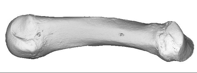
Ultrasound Propagation Through BoneShown below is a 3D-rendered image of a human finger bone using micro-computed tomography (micro-CT).
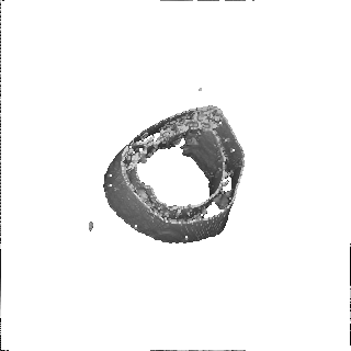
Another example demonstrates propagation through trabecular bone taken from the human heel bone, shown below. This image data was also acquired using micro-CT. The core is approximately 1 cm in diameter and 1.2 cm in length. The propagation is carried out again with a 1 MHz broadband pulse, and the source is placed on the face at one end of the cylinder. Blood is assumed to fill all of the pore spaces and to surround the core as well.
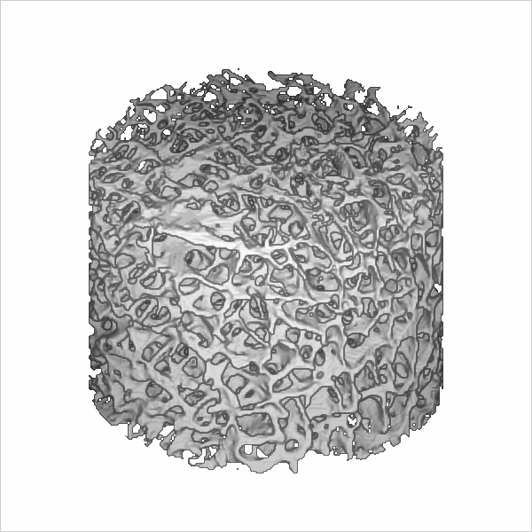
An animated graphic of the propagating wave is shown in the figure below. (Note that the core has been rotated so that the left face is now on the left side.) As may be seen the wave appears to be scattered greatly by the trabeculae. The images of the propagating wave seem to be an "inverse" image of the bone (i.e., the pore spaces); this is likely due to the fact that the displacements are much larger in the pores (fluid) relative to the displacements in the bone material per se.

|
|||||||||
© 2024 CyberLogic, inc. All Rights Reserved