

UltraScan™ 650 ‒‒ The only ultrasound bone densitometer to replace DXA for measuring BMD
|
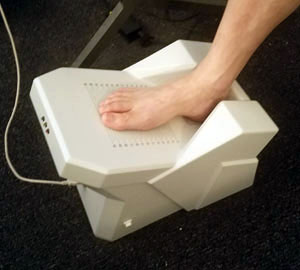
QRT® 250 Heel Scanner
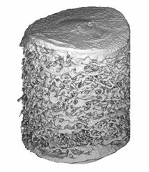
The QRT® 250 Heel Scanner is an ultrasound based device designed to quantitatively assess the calcaneus (heel bone) at a location which is primarily trabecular (~90-96 percent). The calcaneus bone mass has been measured with x-rays for years and shown to be an excellent proxy for fracture risk. The QRT® 250 makes each radiation-free measurement in about a second, with a reproducibility of 3.1 percent. The QRT® 250 Heel Scanner—with its extremely low cost and excellent precision—should allow for much wider diagnosis and detection of individuals at risk of osteoporotic fracture. The QRT® 250 is a through-transmission system that uses two circular single element transducers, operating at 1.5 MHz. The system is designed to measure the calcaneus at a position that depends on the length of the foot, and the transducers are translated to adapt to variations in foot (and calcaneus) size (U.S. and China Patents). The QRT® 250 incorporates CyberLogic's Net Time Delay (NTD) technology (U.S.,China and India Patents). The NTD parameter has been shown to be an excellent proxy for bone mass; see for example the papers located at our Website "A Portable Real-Time Ultrasonic Bone Densitometer" and "Ultrasound Simulation in Bone". These papers describe simulation data as well as clinical data obtained with two earlier versions of the QRT® 250. Using CyberLogic's software package for ultrasound simulation (Wave3000™), we have shown how the NTD technology enables the bone mass, as represented by bone volume fraction, to be accurately estimated. For example, the figure below shows a 3D micro-CT image of a core of a calcaneus.
Thirty (30) such images served in a study of ultrasound propagation using 3D simulations (Wave3000, CyberLogic, Inc.). The simulated received waveforms were processed to obtain the NTD associated with each calcaneal image, and plotted versus the BMD. The BMD was computed directly from the 3D micro-CT image, using an assumed constant tissue density (of 1.85 g.cm-3). As may be seen, there is excellent correspondence between the NTD and BMD in this simulation study (this data has an R-squared value of 0.98).
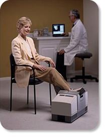
A clinical study was also carried out with an early handheld version of the QRT® 250. Briefly, eighty-five (85) female adult subjects were ultrasonically interrogated and also measured with dual-energy x-ray absorptiometry (GE/Lunar PIXI bone densitometer) at the heel (below).
A linear multivariate regression (using age and NTD as independent variables) was used to determine an estimate of bone mass at the heel. The relationship between the ultrasound-based estimate of heel BMD and actual (DXA-determined) BMD is shown below, and demonstrates a linear correlation of 0.88 (P<0.001). This correlation is higher than presently FDA-approved ultrasound bone assessment devices, which have a correlation under R=0.8.
QRT® 250 Heel Scanner is an investigational device. Limited by US Federal law to investigational use.
|
© 2024 CyberLogic, inc. All Rights Reserved